42 draw and label the microscope
Microscope Drawing Easy with Label - YouTube In this video I go over a microscope drawing that is easy with label. There is a blank copy at the end of the video to review on your own. A great way to s... A Study of the Microscope and its Functions With a Labeled Diagram ... A Study of the Microscope and its Functions With a Labeled Diagram To better understand the structure and function of a microscope, we need to take a look at the labeled microscope diagrams of the compound and electron microscope. These diagrams clearly explain the functioning of the microscopes along with their respective parts. M mooketsi
Core practical 2: Use a light microscope to observe and measure ... microscope: warn against being vigorous with slides as they can splinter and advise on safe use of mounting needles. Practical techniques 4, 5 CPAC 1a, 4a Procedure 1. Before starting this core practical, familiarise yourself with the parts of the microscope and how to focus. Check that the lens and eyepiece are clean, using lens tissue to clean them if necessary as normal tissues or …

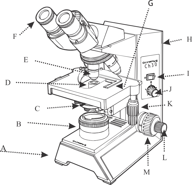
Draw and label the microscope
Microscope, Microscope Parts, Labeled Diagram, and Functions Microscopes magnify or enlarge small objects such as cells, microbes, bacteria, viruses, microorganisms etc. at a viewable scale for examination and analysis. Microscopes consist of one or more magnification lenses to enlarge the image of the microscopic objects placed in the focal plane. Virtual Microscope - NCBioNetwork.org Lesson Description BioNetwork’s Virtual Microscope is the first fully interactive 3D scope - it’s a great practice tool to prepare you for working in a science lab. Explore topics on usage, care, terminology and then interact with a fully functional, virtual microscope. When you are ready, challenge your knowledge in the testing section to see what you have learned. A Study of the Microscope and its Functions With a Labeled Diagram A Study of the Microscope and its Functions With a Labeled Diagram To better understand the structure and function of a microscope, we need to take a look at the labeled microscope diagrams of the compound and electron microscope. These diagrams clearly explain the functioning of the microscopes along with their respective parts.
Draw and label the microscope. Pond Water Under the Microscope Here, students can sketch down what they observe and later label the different parts of the organisms. Conclusion. The primary goal here is for students to observe for themselves the different types of small organisms, which live in the pond and their diversity. Making rough sketches allows them to draw what they see and how they see them. Microscope Labeling Diagram | Quizlet Unit 2 Lesson 5 - Punnett Squares and Pedigrees. 4 terms. PGFry210. Unit 2 Lesson 4 - Heredity. 9 terms. PGFry210. Upgrade to remove ads. Only $2.99/month. Label Microscope Diagram - EnchantedLearning.com Using the terms listed below, label the microscope diagram. arm - this attaches the eyepiece and body tube to the base. base - this supports the microscope. body tube - the tube that supports the eyepiece. coarse focus adjustment - a knob that makes large adjustments to the focus. diaphragm - an adjustable opening under the stage, allowing ... PDF Microscope Practice Actual Size and Drawing Magnification Lab Draw what the object looks like under the microscope. Make sure you get the size of the object compared to the size of the field correct! 3. Label the Name of the object and the Microscope Magnification (Total) beside your drawing. At this point, leave spaces for "Actual Size" & "Drawing Magnification" blank!
Microscope Parts, Function, & Labeled Diagram - slidingmotion Microscope parts labeled diagram gives us all the information about its parts and their position in the microscope. Microscope Parts Labeled Diagram The principle of the Microscope gives you an exact reason to use it. It works on the 3 principles. Magnification Resolving Power Numerical Aperture. Parts of Microscope Head Base Arm Eyepiece Lens Compound Microscope Parts, Functions, and Labeled Diagram Common compound microscope parts include: Eyepiece (ocular lens) with or without Pointer: The part that is looked through at the top of the compound microscope. Eyepieces typically have a magnification between 5x & 30x. Monocular or Binocular Head: Structural support that holds & connects the eyepieces to the objective lenses. Compound Microscope- Definition, Labeled Diagram, Principle, Parts, Uses The naked eye can now view the specimen at magnification 400 times greater and so microscopic details are revealed. Alternatively, the magnification of the compound microscope is given by: m = D/ fo * L/fe where, D = Least distance of distinct vision (25 cm) L = Length of the microscope tube fo = Focal length of the objective lens PDF Label and Color the Microscope Parts - White Plains Public Schools Color the stage dark blue. Draw orange stripes on the body tube Color the diaphragm light green. Color the eyepiece black. Color the stage clips purple. Color the nosepiece light blue. Color the light source yellow Draw green stripes on the inclination joint Draw blue stripes on the coarse focus adjustment knob Color the low-power objective orange
Labeling the Parts of the Microscope | Microscope activity, Science ... Description Worksheet identifying the parts of the compound light microscope. Answer key: 1. Body tube 2. Revolving nosepiece 3. Low power objective 4. Medium power objective 5. High power objective 6. Stage clips 7. Diaphragm 8. Light source 9. Eyepiece 10. Arm 11. Stage 12. Coarse adjustment knob 13. Fine adjustment knob 14. Base How to draw compound of Microscope easily - step by step I will show you " How to draw compound of microscope easily - step by step "Please watch carefully and try this okay.Thanks for watching.....#microscopedrawi... Parts of a microscope with functions and labeled diagram Ans. Microscopes are instruments that are used in science laboratories, to visualize very minute objects such as cells, and microorganisms, giving a contrasting image, that is magnified. Q. State functions of a microscope. Ans. A microscope is usually used for the study of microscopic algae, fungi, and biological specimens. How to draw and label the parts of a microscope? What are at least 3 ... Answer (1 of 3): Google images of light microscope, phase contrast microscope, fluorescent microscope, transmission electron microscope, scanning electron microscope. Label eyepiece, phototube, objectives, stage, sub stage condenser, light (electron) source, focusing knobs, and microscope base. ...
Parts of the Microscope with Labeling (also Free Printouts) Parts of the Microscope with Labeling (also Free Printouts) A microscope is one of the invaluable tools in the laboratory setting. It is used to observe things that cannot be seen by the naked eye. Table of Contents 1. Eyepiece 2. Body tube/Head 3. Turret/Nose piece 4. Objective lenses 5. Knobs (fine and coarse) 6. Stage and stage clips 7. Aperture
Label the microscope — Science Learning Hub Drag and drop the text labels onto the microscope diagram. If you want to redo an answer, click on the box and the answer will go back to the top so you can move it to another box. If you want to check your answers, use the Reset incorrect button. This will reset incorrect answers only.
Microscope Parts and Functions First, the purpose of a microscope is to magnify a small object or to magnify the fine details of a larger object in order to examine minute specimens that cannot be seen by the naked eye. Here are the important compound microscope parts... Eyepiece: The lens the viewer looks through to see the specimen.
Labeling the Parts of the Microscope Labeling the Parts of the Microscope This activity has been designed for use in homes and schools. Each microscope layout (both blank and the version with answers) are available as PDF downloads. You can view a more in-depth review of each part of the microscope here. Download the Label the Parts of the Microscope PDF printable version here.
Simple Microscope - Parts, Functions, Diagram and Labelling Stage - The stage of the microscope is a metal plate that is rectangular in shape and fitted to the vertical rod. It comes with a hole in the center that enables the light to pass from below. The stage holds the slide that contains the specimen to be examined for.
Parts of a microscope with functions and labeled diagram 19.04.2022 · Thank you very much it really helped me with my science home work since i in 8th grade and this my home work to draw a microscope label all the parts and the function thank may the holy father of holy spirits bless you and give more wisdom thanks love you all keep up the good work and thank you again bye. Reply . Sagar Aryal. May 21, 2022 at 1:57 AM . Thank you …
PDF Label parts of the Microscope: Answers Label parts of the Microscope: Answers Coarse Focus Fine Focus Eyepiece Arm Rack Stop Stage Clip . Created Date: 20150715115425Z ...
PRACTICAL BOOKLET - BIOLOGY4ISC Draw borderlines on all four sides of 1 cm on the blank page. Explanation, result or inference to be written on the ruled page. Observation table for physiology experiments should be on the blank page. In taxonomy experiment, classification (for study of flowers) to be written on Right Hand Side corner of the blank page. Draw the diagrams in the middle of the page in such a way that …
Microscope Labeling - The Biology Corner Students label the parts of the microscope in this photo of a basic laboratory light microscope. Can be used for practice or as a quiz. ... Microscope Labeling . Microscope Use: 15. When focusing a specimen, you should always start with the _____ objective. 16. When using the high power objective, only the _____ knob should be used. 17. The ...


Post a Comment for "42 draw and label the microscope"