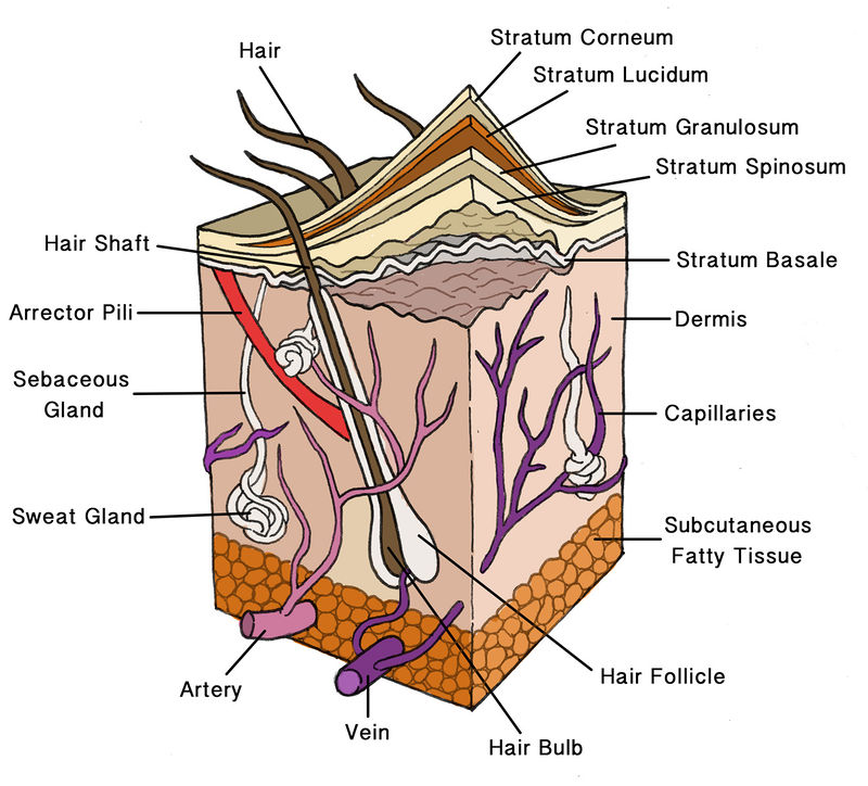
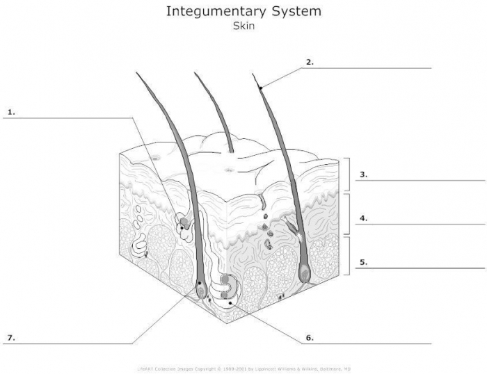
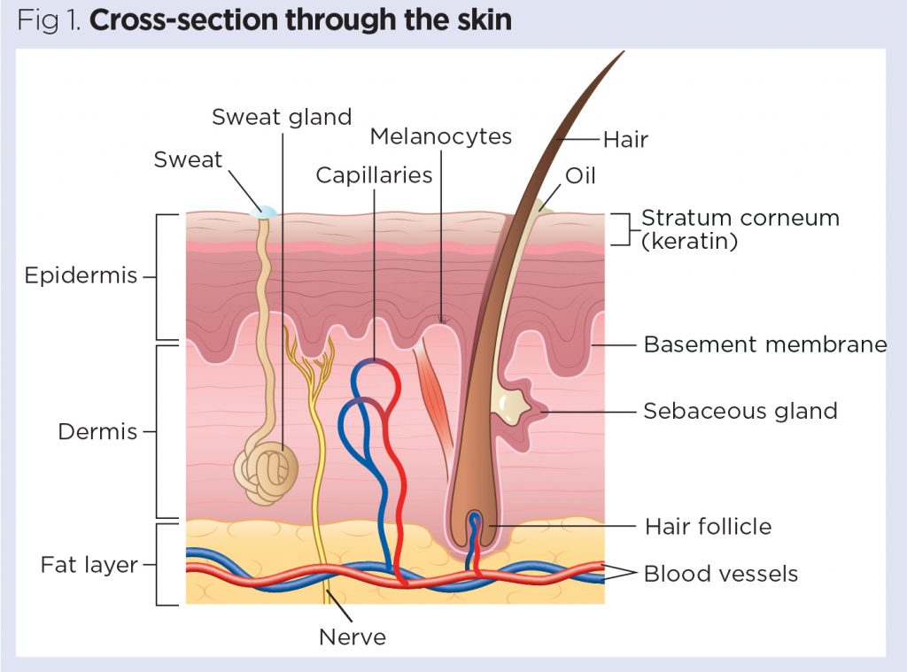
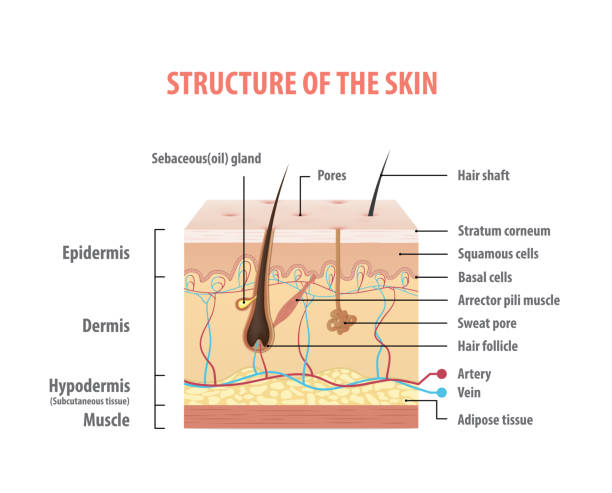
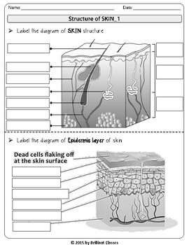
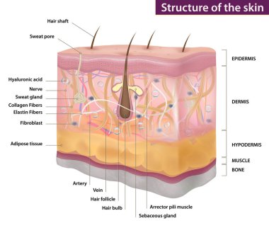
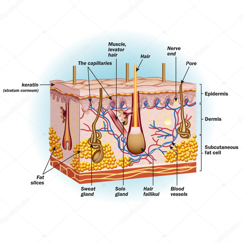
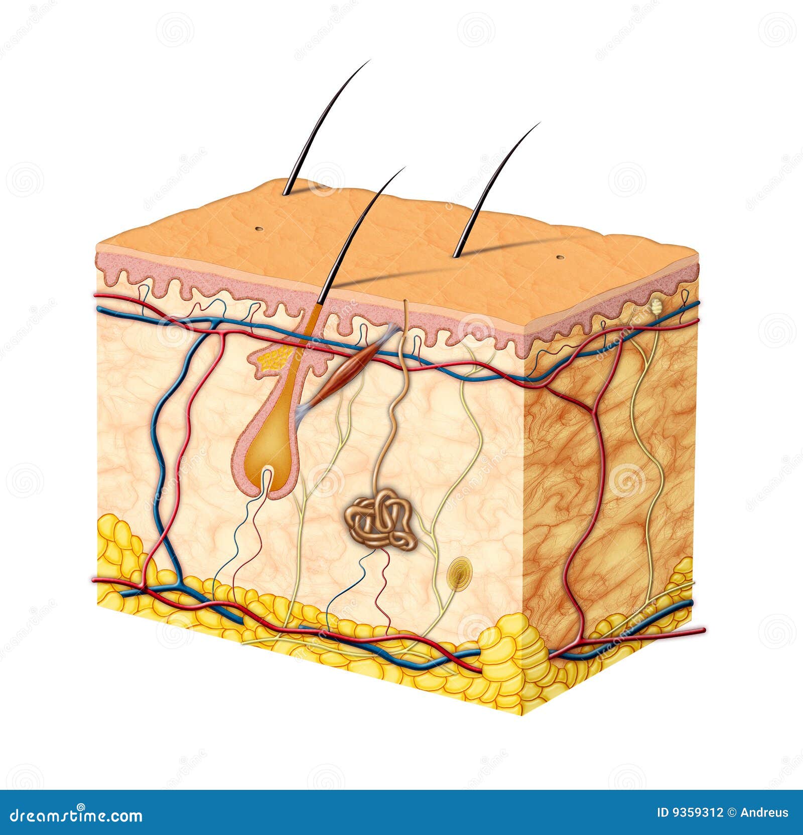
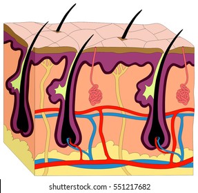
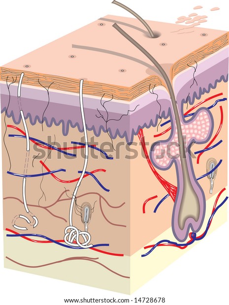
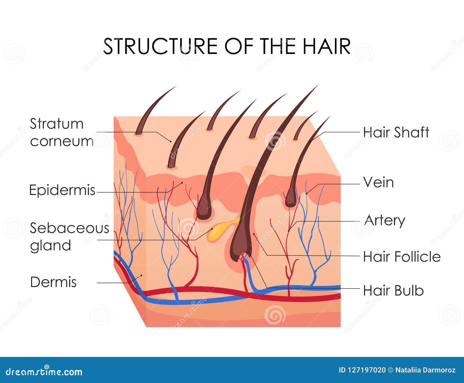
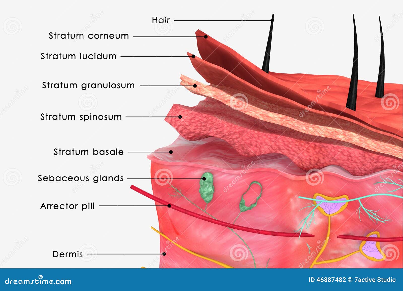
40 label skin diagram
Learn More About Different Body Parts Of Chicken - ROYS FARM We can also determine healthy and unhealthy chicken by following the body parts of chicken. Body Parts Of Chicken Comb Nostril Beak Eye Ear Lobe Wattle Wing Tail Vent Leg Body Parts Of Chicken Small description of different important body parts of chicken are described below. Comb Comb is a small ice of meat above the chickens head. › articles › what-areDermatomes: Definition, chart, and diagram - Medical News Today Apr 30, 2020 · A dermatome is an area of skin that sends information to the brain via a single spinal nerve. Spinal nerves exit the spine in pairs. There are 31 pairs in total, and 30 of these have corresponding ...
Linezolid - Wikipedia Linezolid is an antibiotic used for the treatment of infections caused by Gram-positive bacteria that are resistant to other antibiotics. Linezolid is active against most Gram-positive bacteria that cause disease, including streptococci, vancomycin-resistant enterococci (VRE), and methicillin-resistant Staphylococcus aureus (MRSA). The main uses are infections of the skin and pneumonia ...

Label skin diagram
Penis: Anatomy, Function, Disorders, and Diagnosis - Verywell Health Glans: The glans, or head of the penis, is the sensitive structure at the end of the corpus (shaft).; Urethra: The urethra is a tube inside the penis that runs from the bladder to the head of the penis. It is used for urination. It also crosses through the prostate gland, where an opening (called the ejaculatory duct) receives sperm and fluids that mix together to form semen. Human Skin Diagram Unlabeled - human anatomy review and question ... Human Skin Diagram Unlabeled - 18 images - identifying information from a graphic representation of information, skin anatomy diagram description illustration skin stock illustration, section of human skin, human skin and hair folicle vector stock vector illustration of root, plantuml.com › object-diagramObject Diagram syntax and features - PlantUML.com PlantUML object diagram syntax: You can define objects, fields, relationships, packages, notes... Changing fonts and colors is also possible.
Label skin diagram. Image Classifier using CNN - GeeksforGeeks Since it's not an article explaining CNN so I'll add some links in the end if you guys are interested in how CNN works and behaves. So after going through all those links let us see how to create our very own cat-vs-dog image classifier. For the dataset we will use the Kaggle dataset of cat-vs-dog: train dataset- link. test dataset- link. Integumentary system: Definition, diagram and function | Kenhub The skin is the largest component of this system. It is an extensive sensory organ, which forms an outer, protective coat around the entire external surface of the body. In fact, it is the largest organ of the human body, covering an area of 2 square meters. It has a thickness between 1.5 and 5 mm, depending on location. Functions › game › 1566d27ff4Skin Labeling Quiz - PurposeGames.com Oct 07, 2009 · This is an online quiz called Skin Labeling. There is a printable worksheet available for download here so you can take the quiz with pen and paper. From the quiz author. Manage sensitivity labels in Office apps - Microsoft Purview ... In this article. Microsoft 365 licensing guidance for security & compliance.. When you have published sensitivity labels from the Microsoft Purview compliance portal, they start to appear in Office apps for users to classify and protect data as it's created or edited.. Use the information in this article to help you successfully manage sensitivity labels in Office apps.
Trunk Region (Torso) < Regional Anatomy - Wellness Advocate . com Breasts In our body's trunk region, the paired Mammary Region (Breasts), in the front of the chest encompassing each breast and housing the mammary glands, composed mainly of skin, muscles, fat (adipose tissue), breast tissue and connective tissue, nerves, veins, and arteries. In and on our body's chest, each Breast contains 15 to 20 lobes arranged in a circular fashion. What Required Information Must GHS Labels Include? - MPC For instance, all shipped hazardous chemical containers must be labeled with a signal word, pictogram, hazard statement, and a precautionary statement for each hazard class and category. These requirements impact chemical manufacturers, importers, and distributors. More than 65 countries are using the GHS system or are currently in the process ... Structure of the Skin: Cross-section through the Skin, Diagrams Structure of the Skin: Skin is the largest organ of our body that forms the protective covering of our body. It serves as a barrier between our body organs and the external environment. Beyond providing protection, it plays a crucial role in regulating body temperature. It possesses thermoreceptors and thereby serves as a sense organ. 30 Informative Facts, Diagram & Parts Of Human Body For Kids - MomJunction The human blood is composed of red blood cells, white blood cells, platelets, and plasma. Up to 60% of the human body is water. A human heart beats around 100,000 times in a day. An adult heart beats around 60-80 times per minute, while a child's heart beats 100 - 120 times. Skin is the largest human organ.
Gram Stain Technique - Amrita Vishwa Vidyapeetham Part 2: Labeling of the slides Drawing a circle on the underside of the slide using a glassware-marking pen may be helpful to clearly designate the area in which you will prepare the smear. You may also label the slide with the initials of the name of the organism on the edge of the slide. Layers Of Skin Integumentary System Quiz - ProProfs Quiz 1. Made up of the membrane sheet called integument. A. Dermis B. Integumentary System C. Epidermal layer D. Stratum germinativum 2. What are the appendages of the Integumentary system? A. Hair, nails, skin glands B. Hair, eye color, nails C. Nails, skin color, hair D. Legs, arms, toes 3. What are the two layers of the skin? A. Stratum Germinativum Positions and Functions of the Four Brain Lobes | MD-Health.com The occipital lobe, the smallest of the four lobes of the brain, is located near the posterior region of the cerebral cortex, near the back of the skull. The occipital lobe is the primary visual processing center of the brain. Here are some other functions of the occipital lobe: Visual-spatial processing. Movement and color recognition. Anatomy of the spinal cord - e-Anatomy - IMAIOS This atlas of human anatomy describes the spinal cord through 18 anatomical diagrams with 270 anatomical structures labeled. It was designed particularly for physiotherapists, osteopaths, rheumatologists, neurosurgeons, orthopedic surgeons and general practitioners, especially for the study and understanding of medullary diseases.
art labeling activity the structure of the epidermis - pinklipspro Structure of the epidermis Part A Drag the appropriate labels to their respective targets. A finger-like projection or fold known as the dermal papilla. Start studying Art-labeling Activity. 8 rows Art-labeling activity. Label the diagram by placing the correct letter in the box a.
Pepper Anatomy: What's Inside Your Chili? - PepperScale Pericarp. The pericarp is the whole wall of the fruit. It refers to everything that encloses the fruit's interior. The pericarp consists of three layers. The shiny, waxy outermost layer of the chili pepper is called the exocarp. The exocarp's function is primarily protective. Underneath the exocarp is a middle wall called the mesocarp.
› science › biology-diagramFree Biology Diagram Software with Free Templates - EdrawMax File embedment: Embed the biology diagram on different email attachments and website links and let others benefit from it. Presentation mode: Drag the cursor to the complex part of the biology diagram and zoom it to emphasize any diagram part. Click F5 to enable the full-screen mode to deliver a clear presentation.
Hypodermis of the Skin Anatomy and Physiology - Verywell Health Your skin provides a barrier against fluids, infection, and trauma. It is the largest organ in your body and is made of three different layers: the epidermis (outer layer), the dermis (middle layer), and the hypodermis (bottom or inner layer). 1 The hypodermis is also known as the subcutaneous layer or subcutaneous tissue.
en.wikipedia.org › wiki › MelanomaMelanoma - Wikipedia Melanoma, also redundantly known as malignant melanoma, is a type of skin cancer that develops from the pigment-producing cells known as melanocytes. Melanomas typically occur in the skin, but may rarely occur in the mouth, intestines, or eye (uveal melanoma).
How to Work Safely with - Hazardous Products using the "Skull and ... What does this pictogram mean? The symbol within the pictogram is a human skull with two crossed bones behind it. The symbol indicates that hazardous products with this pictogram can cause death or poisoning. Hazardous products with this pictogram can be safely worked with if proper storage and handling practices are followed.
Understanding an ECG | ECG Interpretation | Geeky Medics An ECG electrode is a conductive pad that is attached to the skin to record electrical activity. An ECG lead is a graphical representation of the heart's electrical activity which is calculated by analysing data from several ECG electrodes. A 12-lead ECG records 12 leads, producing 12 separate graphs on a piece of ECG paper.
Organizational Chart Examples for Any Organization - Creately Blog Organization Structure Template for Corporate Business. Often, the organizational structure can be large in size. Hence it looks more complex to understand. The matrix organizational chart is often used when you have personal reporting to multiple teams or departments. Refer the previously linked article about different types of organizational ...
Anatomy and Structure of the Human Eye (With Diagrams) The retina is the innermost layer lining the back of the eyeball and the light-sensitive part of the eye. The retina contains photoreceptors that detect light. These photoreceptors are known as cones and rods. Cones enable us to detect colours, while rods help us to see in poor light. The retina contains nerve cells that transmit signals from ...
WHMIS 2015 - Pictograms : OSH Answers - Canadian Centre for ... Most pictograms have a distinctive red "square set on one of its points" border. Inside this border is a symbol that represents the potential hazard (e.g., fire, health hazard, corrosive, etc.). Together, the symbol and the border are referred to as a pictogram. Pictograms are assigned to specific hazard classes or categories.
remm.hhs.gov › ext_contaminationProcedures for Radiation Decontamination - Radiation ... Update body diagram after each decontamination cycle or use new body diagram for each cycle. Begin decontamination with areas of highest contamination. In a water deficient environment, gently brush skin surface to remove a portion of the stratum corneum layer and dislodge contamination held by skin proteins.
Skin: Cells, layers and histological features | Kenhub The epidermis is the uppermost layer of the skin. Going from deep to superficial, it consists of five layers; basal layer (stratum basale/germinativum) prickle cell layer (stratum spinosum) granular layer (stratum granulosum) clear layer (stratum lucidum) cornified layer (stratum corneum) To remember these layers, check out this mnemonics video:
How to: Create a Drill-Down Chart - DevExpress To add chart titles, select the chart, and click the ellipsis button for its ChartControl.Titles property. In the invoked Chart Title Collection Editor, click Add, to create a title. Set its Title.Text property to "Back to the main view…", and the TitleBase.Visible property to false. Add another chart title ( with the "Sales by Person ...
Stress-Strain Curve: Definition, Formula, Hooke's Law - Embibe On plotting the stress and its corresponding strain on the graph, we get a curve, and this curve is called the stress-strain curve or stress-strain diagram. Generally, we take the strain (no units) on the \ (x-\)axis and stress (\ (\rm {Pa}\) or \ (\rm {MPa}\)) on the \ (y-\)axis.
Structure and parts of a sperm cell - inviTRA This labelled diagram shows the structure of a sperm cell in detail, which has the following parts: Head With its spheric shape, it consists of a large nucleus, which at the same time contains an acrosome. The nucleus contains the genetic information and 23 chromosomes. It also secretes a hyaluronidase enzyme that destroys the hyaluronic acid ...
checkify.com › blog › fishbone-diagramFishbone Diagram: Determine Cause and Effect • Checkify You can have major causes as the main branches while minor ones form the sub-branches. The diagram can have as many branches as the project requires. Identifying all the reasons for the problems faced allows managers to search for the best solutions. Fishbone Diagram History. Fishbone diagram was created by Dr. Kaoru Ishikawa (1915-1989)
How to Remove Skin Tags | U.S. News Apply the tea tree oil or vinegar to the skin tag and massage it into the surface. Cover the area with a bandage. With tea tree oil, leave the bandage in place overnight, but if you're using...
Human Biology Lab Online | Lab 4 Tissues and Skin Select the Labels "On" button Use the slider bars on the side and bottom to scan around the slide. View the tissue at 1000x to see the fine ultra structure Note: Browser Pop-up Blockers must be turn off to view these slides. Click on any of the slides listed above on the tissue slide box image to view.
plantuml.com › object-diagramObject Diagram syntax and features - PlantUML.com PlantUML object diagram syntax: You can define objects, fields, relationships, packages, notes... Changing fonts and colors is also possible.
Human Skin Diagram Unlabeled - human anatomy review and question ... Human Skin Diagram Unlabeled - 18 images - identifying information from a graphic representation of information, skin anatomy diagram description illustration skin stock illustration, section of human skin, human skin and hair folicle vector stock vector illustration of root,
Penis: Anatomy, Function, Disorders, and Diagnosis - Verywell Health Glans: The glans, or head of the penis, is the sensitive structure at the end of the corpus (shaft).; Urethra: The urethra is a tube inside the penis that runs from the bladder to the head of the penis. It is used for urination. It also crosses through the prostate gland, where an opening (called the ejaculatory duct) receives sperm and fluids that mix together to form semen.










Post a Comment for "40 label skin diagram"