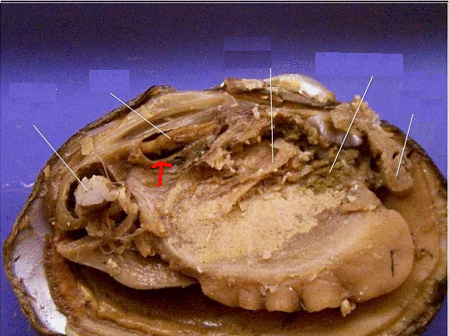
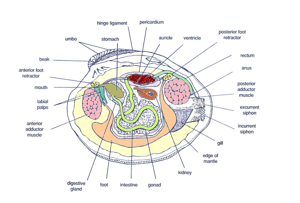
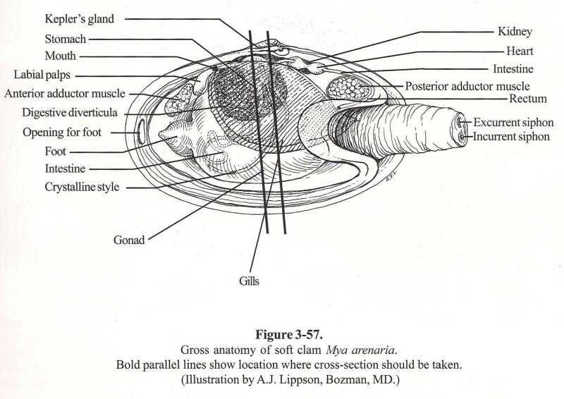
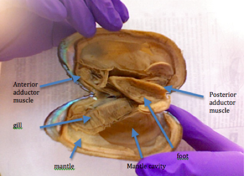
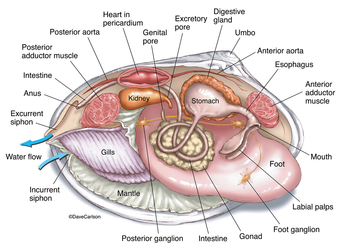
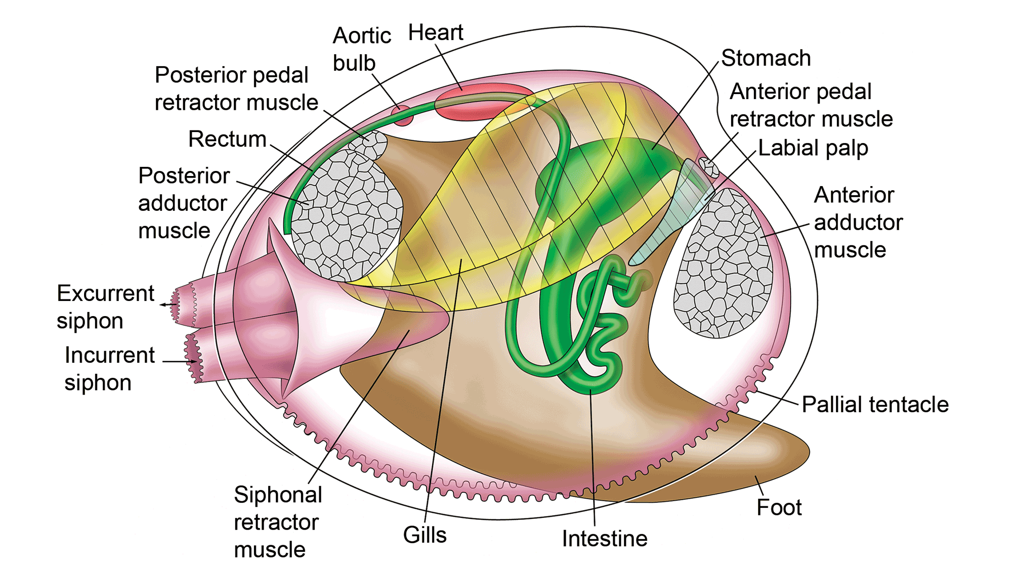
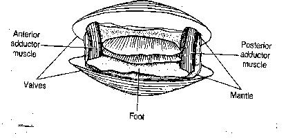
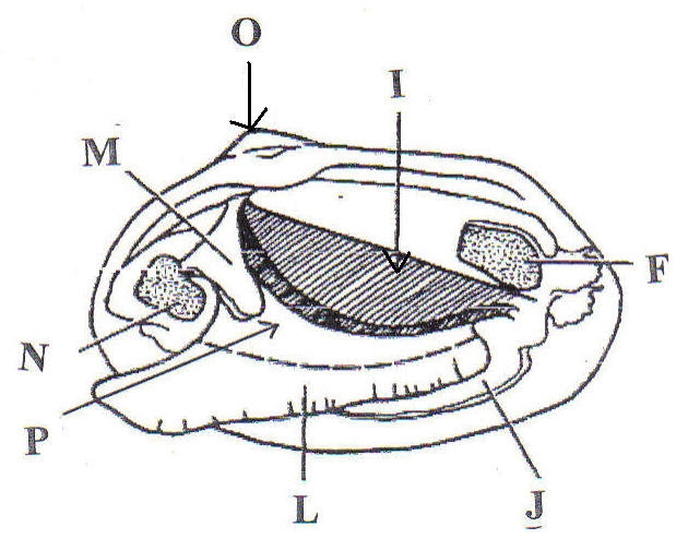
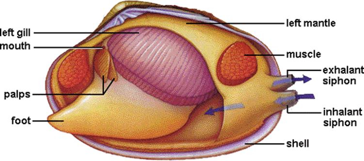
43 internal structure of a clam labeled
PDF Anatomy of a Clam - University of Florida Obtain clam specimens (fresh or preserved). 2. Divide the class into small groups (2-4 per group when possible). 3. Prepare one dissection kit, pan, and clean-up materials per group. 4. Copy the dissection guide for each student. 5. Copy the Externaland Internal Clam Anatomyhandouts for each student. 6. PDF Biology 11 Name: Clam Dissection 1 anterior posterior 2. - Mrs Dildy Diagram 2: Clam Internal Anatomy 6. With scissors, cut off the ventral portion of the foot. Use the scalpel to carefully cut the muscle at the top of the foot into right and left halves. 7. Carefully peel away the muscle layer to view the internal organs. 8. Locate the spongy, yellowish reproductive organs. 9.
bivalve - Internal features | Britannica The general classification of the bivalves is typically based on shell structure and hinge and ligament organization. The internal anatomy is also a tool in classification, particularly the organs of the mantle cavity, the pattern of water movement through it, and the structure and functioning of the ctenidia and labial palps. Early anatomists established a correlation between shell and gill ...
Internal structure of a clam labeled
Clam Dissection Clams are marine mollusks with two valves or shells. ... To study the internal and external anatomy of a bivalve mollusk. Materials Mollusk Anatomy | Carlson Stock Art Illustration of the internal structure of a clam. Browse images in the Categories, or enter a search term here to search the image archives. Narrow your search results by separating multiple keywords with a comma. What is a internal structure? - Answers An Internal Structure is the way an organism looks on the outside and an External Structure is the looks on the outside. ... How can you label the internal structure of a clam?
Internal structure of a clam labeled. Clam study: the shell, the internal anatomy and how they feed ... Clam study: the shell, the internal anatomy and how they feed. Summary. Compare different sizes of shells and learn about how shells grow. Dissect a clam and discover that inside a familiar clam shell, often seen on the beach, there is a living animal. Identify the major body parts of a clam, and compare their function to equivalent organs in ... Clam to Label Quiz - PurposeGames.com Soil Profile - Horizon Label and Name 12p Image Quiz. Mushroom Diagram 7p Image Quiz. Cave Deposition 4p Image Quiz. Topographical Map 6p Image Quiz. Feather Structure 6p Image Quiz. Squid to label 17p Image Quiz. Human Body Systems 10p Matching Game. Clam Anatomy - Clams Ahoy The common hardshell clam Mercenaria mercenaria, better known as a cherrystone, has a mouth, labial palps (antecedents of lips), a stomach, separate digestive gland, an intestine, nerve cord and an anus. The foot of a clam is a curved flesh protrusion from the perimeter of the anterior flesh. Clam dissection: A first step into dissection and anatomy for young ... This is a long thin strand of muscle that is often wavy and the part of the clam that you can see when the shell is slightly open. This is known as the dorsal body, and it is like a robe that covers the internal organs of the clam. Over time, the mantle secretes what will become the shell, allowing the animal to grow a larger home. Foot
Anatomy of the Geoduck Clam - Fisheries and Oceans Canada Internal Anatomy The inner surface of the valves (shell) is usually rough with a deeply impressed, continuous pallial line (where the mantle was sealed to the shell) and triangular pallial sinus (Fig. 3). At the dorsal surface, the hinge ligament joins the two valves and each valve has a cardinal tooth at the hinge. Bivalve Anatomy - Paleontological Research Institution The valve structure and soft anatomy of the bivalves varies among species. The Hard-Shelled Clam or Cherrystone, Mercenaria mercenaria, is used here as an example of a typical bivalve. two adductor muscles (reduced to one in some species) that hold the shell tightly closed when they contract. an elastic ligament that springs the shell open when ... PDF Lab 5: Phylum Mollusca - Amherst College Examination of Internal Structure 1. The lab TA's will open a clam for you. Keep the shell that has the soft tissues of the clam attached to it submerged in cold sea water while you examine it (you may get to see the heart beat if you do so!). First, however, examine the other valve. Fig. 3.63. Diagram of the internal anatomy of a clam Fig. 3.63. Diagram of. Fig. 3.63. Diagram of the internal anatomy of a clam. Image by Byron Inouye.
Clam Dissection The tube-like structure that runs through the pericardium is the intestine. 25. Answer the questions on your lab report & label the diagrams of the internal structures of the clam. Also, use arrows on the clam diagram to trace the pathway of food as it travels to the clam's stomach. Continue the arrows showing wastes leaving through the anus. Clam - Wikipedia A clam's shell consists of two (usually equal) valves, which are connected by a hinge joint and a ligament that can be internal or external. The ligament provides tension to bring the valves apart, while one or two adductor muscles can contract to close the valves. Clams also have kidneys, a heart, a mouth, a stomach, and a nervous system. Clam structures and functions Flashcards | Quizlet anterior and posterior muscles open and close the valves incurrent siphon brings in food laden oxygenated water excurrent siphon sends waste and deoxygenated water out gills remove oxygen and food particles from the water; moves water throughout the clam using cilia to create a current through the siphons palps [Solved] Draw a diagram and label the internal anatomy of the clam ... Mollusks abandon a more active life style for a sedentary existence like the clam. Mollusks are mostly marine organisms that dwell in a variety of settings ranging from shallow coastal areas to deep seas. The majority of them live in the sediments at the bottom of bodies of water, however others, such as cephalopods, are free-swimming.
Lynn_LabRpt11.docx - Kingdom Animalia: Protostomes Lab... Clam Structures and Functions Structure Description Valve Shells, supportive exoskeleton, composed of protein, chitin, and calcium carbonate. Hard, brown, in color. Posterior and anterior muscles Thick string like muscle from on valve to the other. Brown and peach in color.
PDF Clam Dissection Information Sheet - Internal Anatomy Clam Dissection Information Sheet -Internal Anatomy A. Feeding Mechanism of clam. Food in mucous string Water enters the mantle cavity from the rear and is pulled forward by the beating of cilia to the gills and mouth.
internal anatomy of a clam Thumb Muscle Anatomy boundbobskryptis.blogspot.com. muscle hand anatomy forearm pediatric. Fresh Water Mussel Collection: Introduction - Anatomy -- Illinois State . mussel mussels anatomy water labeled fresh feeding state freshwater tissues soft. Clam Anatomy Labeling Page . clam labeling anatomy ...
anatomy of clam clam diagram labeled internal structures anatomy01 label incurrent excurrent siphon lab diagrams answer Images For BIO 122 Lab klemow.wilkes.edu unlabeled dissected lab earthworm labeled anterior wilkes dorsal bio Clam Anatomy (Function Quiz) clam Leevonk.com leevonk.com anatomy clam biology LLA BIOLOGY: Squid Anatomy
Clams Anatomy - Barnegat Bay Shellfish The clam's foot is used to dig down into the sand, and a pair of long siphon s that extrude from the clam's mantle out the side of the shell reach up to the water above (only the exit points for the siphons are shown). Note: this image is colored to differentiate internal organs and are not the actual colors of the clam. Clams are filter ...
Clam Diagram & Parts | What Is a Clam? | Study.com Belonging to a diverse group of animals known as bivalves, clams can be identified by the presence of two valves, or shells, joined by a hinge that allows the two shells to open or close. A...
Clam Dissection Information Sheet – Internal Anatomy Page 1. Clam Dissection Information Sheet – Internal Anatomy.
clam internal anatomy - Microsoft clam anatomy clams external diagram flickr foot body fresh water. Strongylocentrotus Spp., Sea Urchin: Model Echinoderm, Facts, Life . urchin sea anatomy internal system geochembio echinoderm morphology strongylocentrotus vascular water body oral facts pole cycle taxonomy game. Clam Dissection - YouTube . clam ...
Clam anatomy functions Flashcards | Quizlet outer shell layer (protein outer coat) nacreous layer shiny inner shell layer, surrounds parasites, forming pearls Prismatic Layer Middle Layer contains calcium carbonate. anterior/posterior adductors used to close the shell mantle secretes calcium carbonate that makes the shell hard visceral mass Contains internal organs foot used for movement
Clam Dissection - BIOLOGY JUNCTION Clams are marine mollusks with two valves or shells. Like all mollusks, a clam has a mantle which surrounds its soft body. It also has a muscular foot which enables the clam to burrow itself in mud or sand. The soft tissue above the foot is called the visceral mass and contains the clam's body organs. Taxonomy Kingdom - Animalia Phylum - Mollusca
Anatomy of a bivalve | Museum of Zoology - University of Cambridge Anatomy of a bivalve. Inside the shell. Bivalve molluscs come in many shapes and sizes, and live in many different ways. However, there are features shared by all bivalves. Bivalve molluscs are completely enclosed by a shell made of two valves hinged at the top. A hinge ligament made of elastic protein joins the two halves of the shell together ...
The internal, soft tissue anatomy of clam | Download Scientific Diagram Mantle: The soft parts of the clam tissues are are covered by the mantle, which is composed of two thin sheaths of tissue, thickened at the edges ( Fig.3) . The two halves of the mantle are...
Clam Dissection Guideline BACKGROUND: Clams are bivalves, meaning that they have shells consisting of two ... the internal and external structures of a mollusk by dissecting a clam.
PDF Taxonomy, Anatomy, and Biology of the Hard Clam Internal Clam 1 Mantle Shell Anatomy • Covers visceral or body mass • Holds in fluid • Secrets new shell 2. Ant. adductor muscle 3. Post adductor musclePost. adductor muscle • Hold valves shut 4. Pericardium cavity • Region covered with thin Region covered with thin, dark membrane • Contains 2-chambered heart and kidney in a fluid-filled sac 5.
Clam Dissection.docx - Google Docs Continue following the intestine toward the posterior end of the clam. Find the anus just behind the posterior adductor muscle. Use your probe to trace the path of food & wastes from the incurrent siphon through the clam to the excurrent siphon. Answer the questions on your lab report & label the diagrams of the internal structures of the clam.
What is a internal structure? - Answers An Internal Structure is the way an organism looks on the outside and an External Structure is the looks on the outside. ... How can you label the internal structure of a clam?
Mollusk Anatomy | Carlson Stock Art Illustration of the internal structure of a clam. Browse images in the Categories, or enter a search term here to search the image archives. Narrow your search results by separating multiple keywords with a comma.
Clam Dissection Clams are marine mollusks with two valves or shells. ... To study the internal and external anatomy of a bivalve mollusk. Materials











Post a Comment for "43 internal structure of a clam labeled"